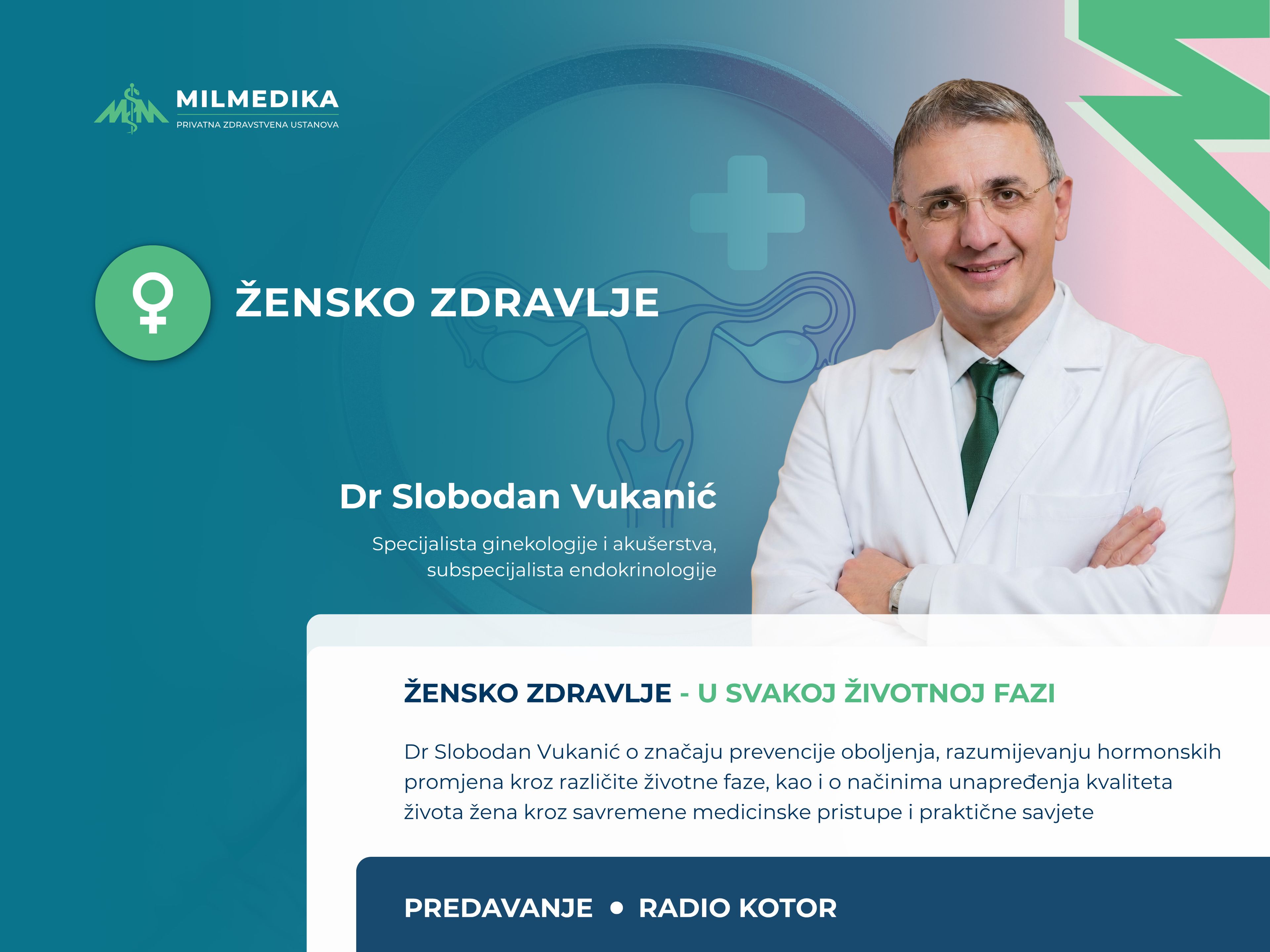
Radiology
Radiology is a vital part of modern medicine, using imaging technologies like ultrasound, X-ray diagnostics, MRI (magnetic resonance imaging), and CT (computed tomography) to examine the inside of the body. These techniques help doctors detect diseases early, track treatment progress, and plan effective therapies.
At Milmedika, we offer ultrasound and X-ray diagnostics with the latest equipment and expert care to provide accurate results and support timely treatment decisions.
Ultrasound diagnostics
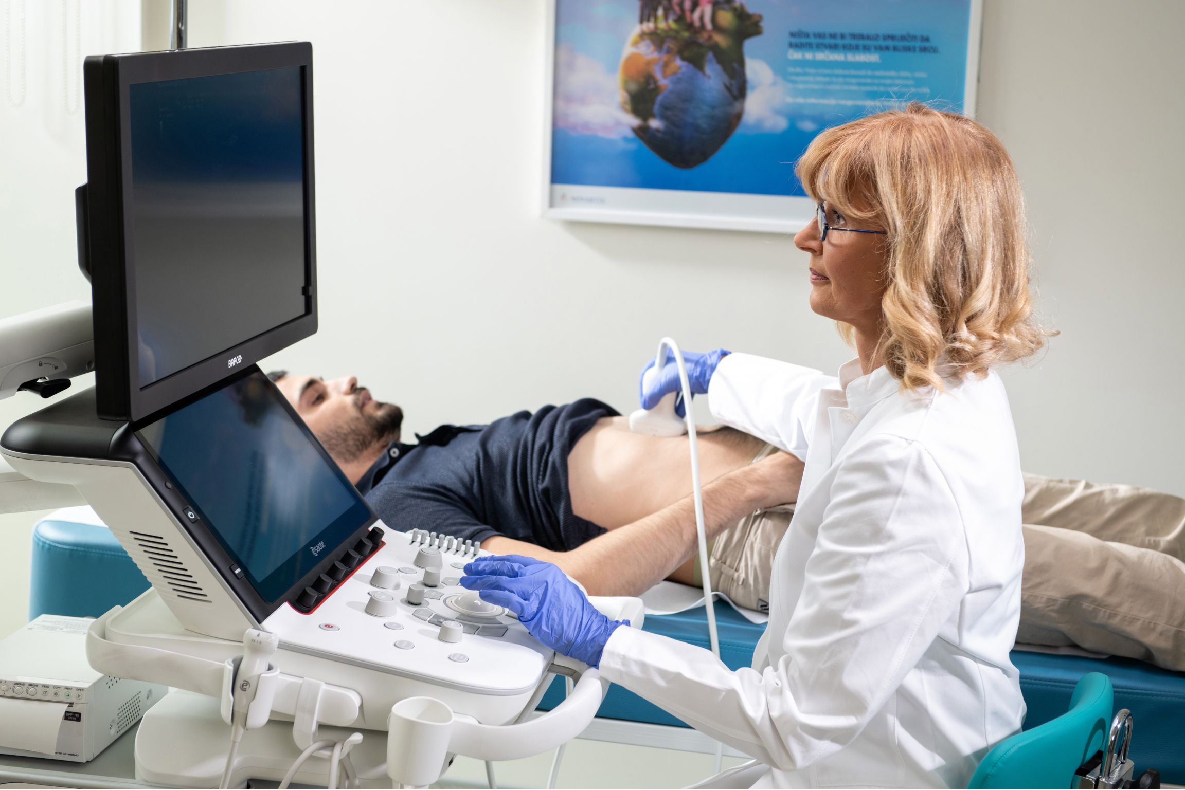
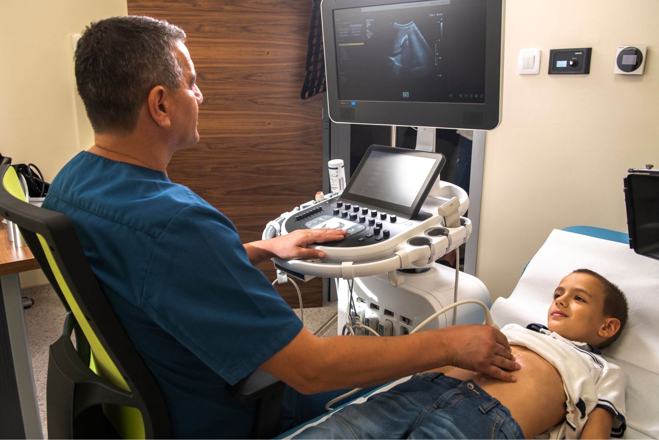
Ultrasound is a safe, painless, and non-invasive diagnostic method that uses sound waves to create clear, real-time images of organs and tissues. Unlike other methods such as X-rays or CT scans, ultrasound doesn’t involve radiation, making it safe for repeated use. It’s a widely used tool for diagnosing many health conditions and is often sufficient for confirming a diagnosis or guiding additional tests.
During the procedure, a small handheld device called a probe is placed on the skin over the area being examined. A special gel is applied to improve contact between the probe and skin, ensuring clear and accurate images. The sound waves from the probe reflect off internal structures, and these reflections are converted into detailed images displayed on a screen.
At Milmedika, we perform ultrasounds using the Philips Affiniti 50 Ultrasound system, a high-performance ultrasound system that provides excellent image quality for precise and reliable diagnostics.
What ultrasound services do we offer?
- Abdominal ultrasound: Examines organs like the liver, gallbladder, pancreas, spleen, kidneys, bladder, and reproductive organs.
- CNS ultrasound: Imaging of the central nervous system.
- Neck ultrasound: Evaluates the thyroid gland, salivary glands, lymph nodes, blood vessels, and neck muscles.
- Breast ultrasound: For screening and evaluating any changes in breast tissue.
- Musculoskeletal ultrasound: Checks muscles, joints, and soft tissues.
- Scrotal ultrasound: Examines the testicles and surrounding tissues.
- Echocardiography: A heart ultrasound to assess its function and structure.
Doppler ultrasound:
This specialized ultrasound method is used to evaluate blood flow through arteries and veins. It is particularly useful for identifying blockages, blood clots, and other vascular issues.
- Doppler of the neck and lower extremities.
- Color Doppler: Assesses blood flow in arteries and veins in the head, neck, heart, abdominal aorta, kidneys, arms, and legs. It can also evaluate blood flow inside the heart chambers.
At Milmedika, our ultrasound diagnostics are designed to detect medical conditions early and guide your care with precision and care, all in a safe and comfortable setting.
X-ray diagnostics
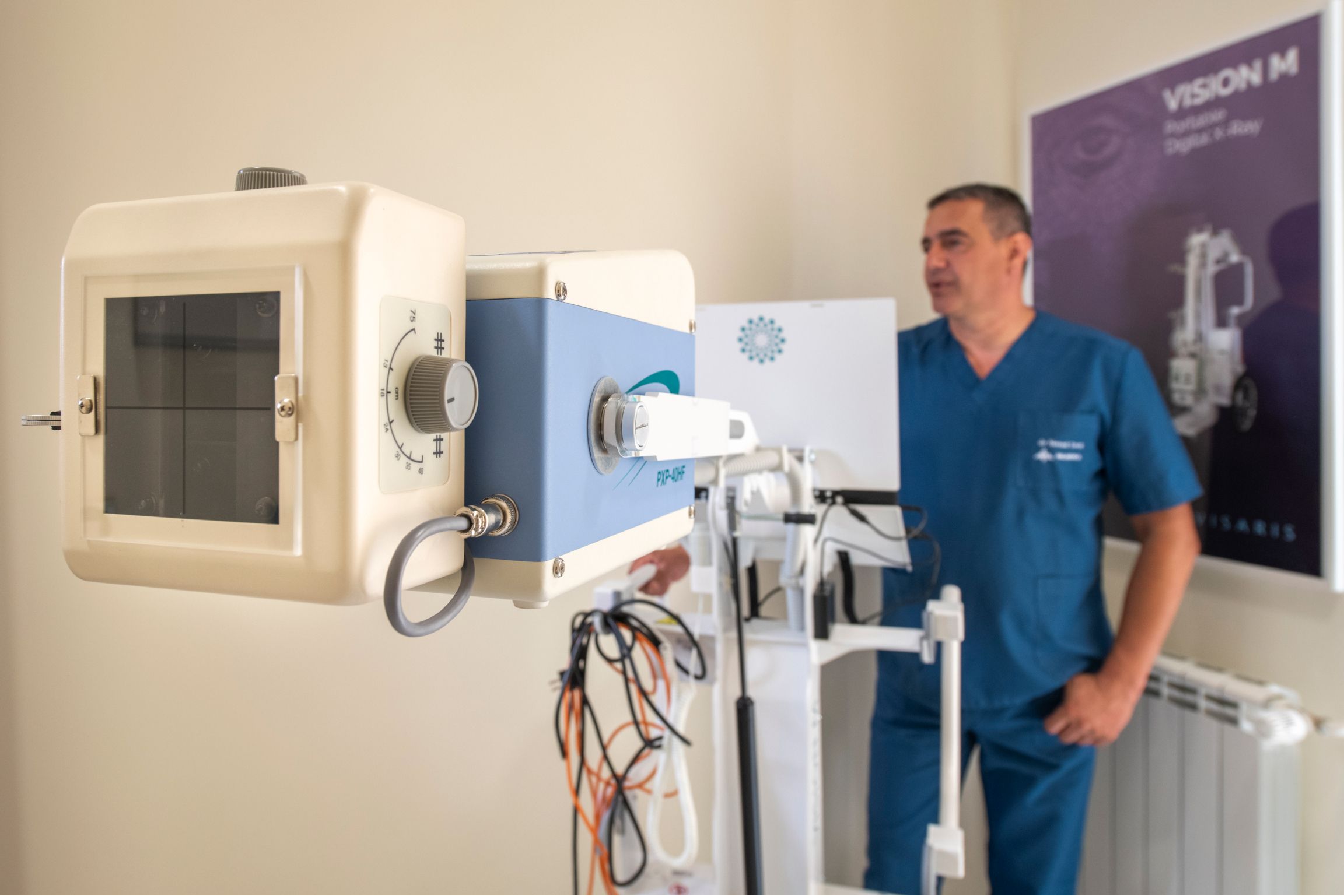
X-rays are one of the most commonly used and reliable diagnostic tools in medicine. They are painless, non-invasive, and quick, producing clear images of the body's internal structures by using a small dose of radiation. X-rays are particularly useful for diagnosing lung conditions, bone and joint problems, and changes in the abdomen or pelvis.
At Milmedika, we use a digital Siemens X-ray system to perform these examinations. This technology ensures high-quality images while minimizing radiation exposure, making it a safe choice for patients of all ages.
How does the procedure work?
Before the procedure, patients are asked to remove clothing or objects (like jewelry, glasses, or watches) that might interfere with the imaging process. Depending on the area being examined, the technician will position you in front of or under the X-ray machine. For some scans, such as a chest X-ray, you may need to hold your breath briefly to prevent any movement that could blur the image.
The imaging itself takes only a few seconds, and the entire procedure usually lasts just a few minutes. Once the images are taken, the radiologist carefully analyzes them and prepares a detailed report explaining the results.
What X-ray services do we offer?
Head and face:
- Skull X-rays (Town, Altschul views).
- Sinus X-rays: Detects blockages or inflammation in the sinuses.
- Orbital (eye socket) X-rays.
- Optic canal X-rays: Used to examine the canal through which the optic nerve passes.
- Mastoid process X-rays: Focuses on the bone behind the ear to check for infections or abnormalities.
- Temporal bone imaging: Examines the structures of the inner ear and temporal bone.
- Pituitary gland (sella turcica) imaging: Evaluates the area where the pituitary gland is located for potential abnormalities.
- Facial bones and nasal imaging: Useful for identifying fractures or other structural issues.
- Jaw and temporomandibular joint (TMJ) imaging: Examines the upper and lower jaw, as well as the jaw joint, to diagnose pain or movement problems.
Spine:
- Cervical (neck) spine: Used to identify conditions like herniated discs or fractures in the neck.
- Thoracic (mid-back) spine: Evaluates issues such as fractures, deformities, or arthritis in the mid-back.
- Lumbar (lower back) spine and sacrum: Commonly used to detect causes of lower back pain or injuries.
- Full spine imaging: Provides a complete view of the spine to assess alignment, scoliosis, or other structural abnormalities.
Joints and extremities:
- Shoulders.
- Upper arms (one or both).
- Elbows (one or both).
- Forearms (one or both).
- Hands (one or both): Often used to detect fractures or joint damage.
- Wrists (one or both).
- Hips.
- Thighs and knees (one or both): Useful for assessing arthritis, fractures, or joint injuries.
- Ankles and feet (one or both): Commonly used to check for sprains, fractures, or bone alignment issues.
- Heels: Examines for bone spurs or other heel-related issues.
Chest and abdomen:
- Lungs and heart: Standard and profile views help assess conditions like pneumonia, lung infections, or heart enlargement.
- Abdomen: Imaging of the abdominal cavity to detect changes in organs such as the intestines or liver.
- Urinary system: Focuses on the kidneys, urinary ducts, and bladder to identify stones, infections, or other issues.
- Pelvis: Useful for assessing fractures, joint conditions, or changes in the reproductive organs.
Why choose digital X-rays?
Digital X-ray technology offers several advantages over traditional film-based systems:
- Lower radiation exposure: Digital X-rays require significantly less radiation, making them safer, especially for children or patients needing multiple scans.
- High-quality imaging: Digital systems provide sharper, more detailed images, helping radiologists make accurate diagnoses.
- Faster results: Digital images are processed immediately, reducing wait times for diagnosis and treatment planning.
At Milmedika, we prioritize your safety and comfort while ensuring you receive the most precise and timely results. X-ray diagnostics play a critical role in identifying and managing health conditions, and our team is here to provide expert care every step of the way.